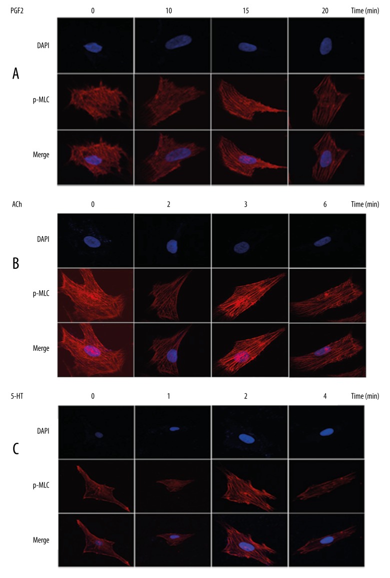
Figure 3.
Increased p-MLC2 level promotes VSMCs contractile activities in vitro. Staining of p-MLC2 using immunofluorescence assay was utilized to reveal myofilaments within the cytoplasm of VSMCs. VSMCs were exposed to PGF2α (A), ACh (B), and 5-HT (C) for indicated times. The best-organized myofilament was observed by exposure to PGF2α for 15 min, to ACh for 3 min, and to 5-HT for 2 min. Extension of exposure time led to dephosphorylation and, hence, less organized myofilament and dimmed fluorescence.

