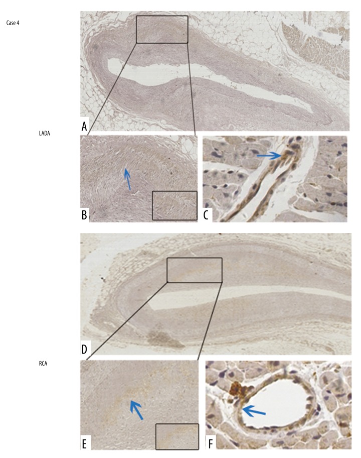
Figure 5.
MLC2 is barely phosphorylated in Case 4. In the LADA, although the lumen was mildly stenosed, p-MLC2 was barely detected under low (A) and high (B) magnification views. Adjacent to the LADA with lesions, the interstitial small artery was also barely stained with p-MLC2 (C). In the RCA, p-MLC2 was barely detected under low (D) and high (E) magnification views. Adjacent to the LADA with lesions, the interstitial small artery was also barely stained with p-MLC2 (F). Magnification: 100× for upper images and 400× for bottom images. Blue arrows indicate mildly positive staining. Magnified insets at right bottoms of (B) and (E) highlight the almost negative staining of p-MLC2 within coronary artery walls.

