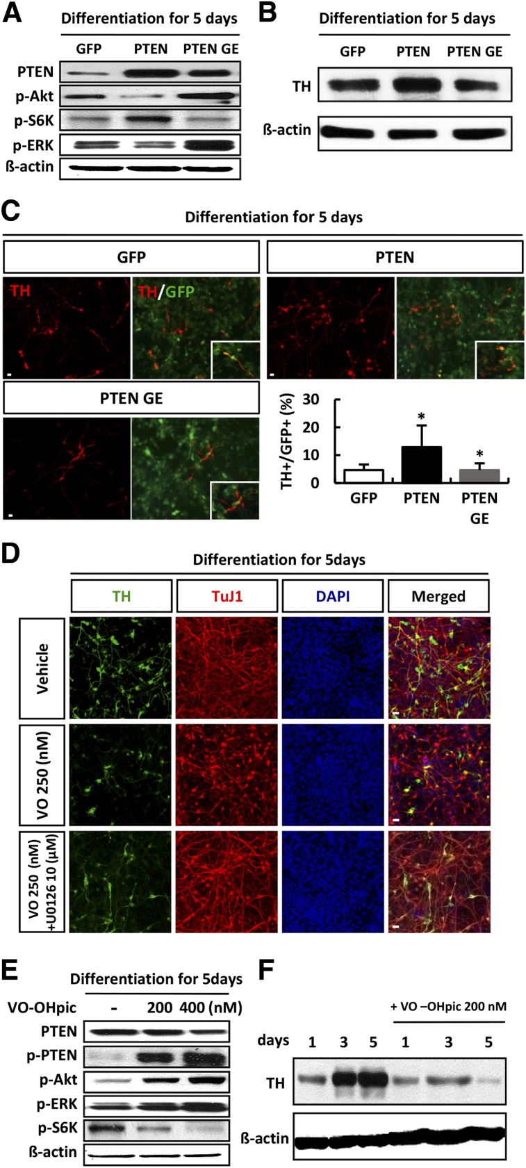
Figure 5.
PTEN promoted dopaminergic neuronal differentiation of human neural stem cells (hNSCs). hNSC cultures were infected in parallel with vectors expressing PTEN-GFP, PTEN G129E-GFP, and GFP. After 3 days, the infected cells were cultured for a further 5 days in conditions of differentiation. (A): Lysates from differentiating cells were subjected to immunoblot analysis of PTEN, p-Akt, p-S6K, p-ERK, and β-actin, as indicated. (B, C): Differentiating cells were subjected to immunoblotting (B) and immunofluorescent staining (C) with anti-TH antibody (red). Green: GFP; blue: DAPI (n = 5). (D–F): Chemical inhibition of PTEN decreases dopaminergic neuronal differentiation. hNSCs were treated with or without VO-OHpic during differentiation (in the absence of basic fibroblast growth factor). Cells positive for TH were quantified as the percentage of GFP-positive cells. (D): Differentiating cells treated with VO-OHpic only or cotreatment with 10 µM U0126 at day 5 were subjected to immunofluorescence with anti-TuJ-1 (red) and anti-TH (green) antibodies; blue indicates DAPI staining. (E): Western blot analysis of PTEN, p-PTEN, p-Akt, p-ERK, p-S6K, and β-actin in hNPCs after treatment with 200 or 400 nM VO-OHpic during differentiation for 5 days. Each Western blot image is representative of three different experiments. (F): Differentiating cells with or without VO-OHpic treatment for 5 days were subjected to immunoblotting for TH and β-actin, as indicated. Error bars are standard errors of the mean (SE). ∗, p < .05, in comparison with control cells. Scale bar = 20 μm. Abbreviations: DAPI, 4′,6-diamidino-2-phenylindole; ERK, extracellular signal-regulated kinase; GFP, green fluorescent protein; p, phosphorylated; PTEN, phosphatase and tension homolog PTEN GE, a catalytically inactive PTEN mutant; S6K, S6 kinase; TH, tyrosine hydroxylase.

