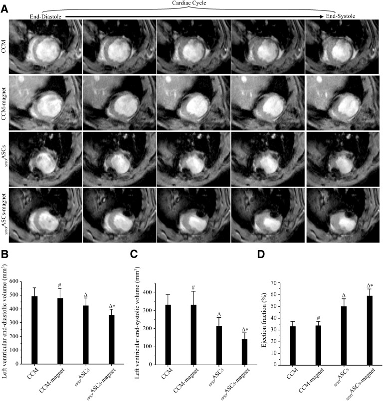
Figure 7.
Magnetic resonance imaging (MRI) analysis of cardiac function. (A): Typical short-axis cine MRIs from end-diastole to end-systole during the whole cardiac cycle. The SPIOASC-magnet-treated rats (n = 14) displayed a smaller left ventricular (LV) slice volume in comparison with the SPIOASC-treated rats (n = 14), the CCM-magnet-treated rats (n = 8), and the CCM control rats (n = 8). (B, C): LV end-diastolic (B) and end-systolic (C) volumes were lowest in the SPIOASC-magnet-treated rats (n = 14) compared with the SPIOASC-treated rats (n = 14), the CCM-magnet rats (n = 8), and the CCM control rats (n = 8). (D): LV ejection fraction was highest in the SPIOASC-magnet-treated rats (n = 14) compared with the SPIOASC-treated rats (n = 14), the CCM-magnet rats (n = 8), and the CCM control rats (n = 8). #, p > .05 versus the CCM; Δ, p < .05 versus the CCM or CCM-magnet; ∗, p < .05 versus the SPIOASCs, CCM-magnet, or CCM. Abbreviations: CCM, cell culture medium; SPIOASC, superparamagnetic iron oxide-labeled adipose-derived stem cell.

