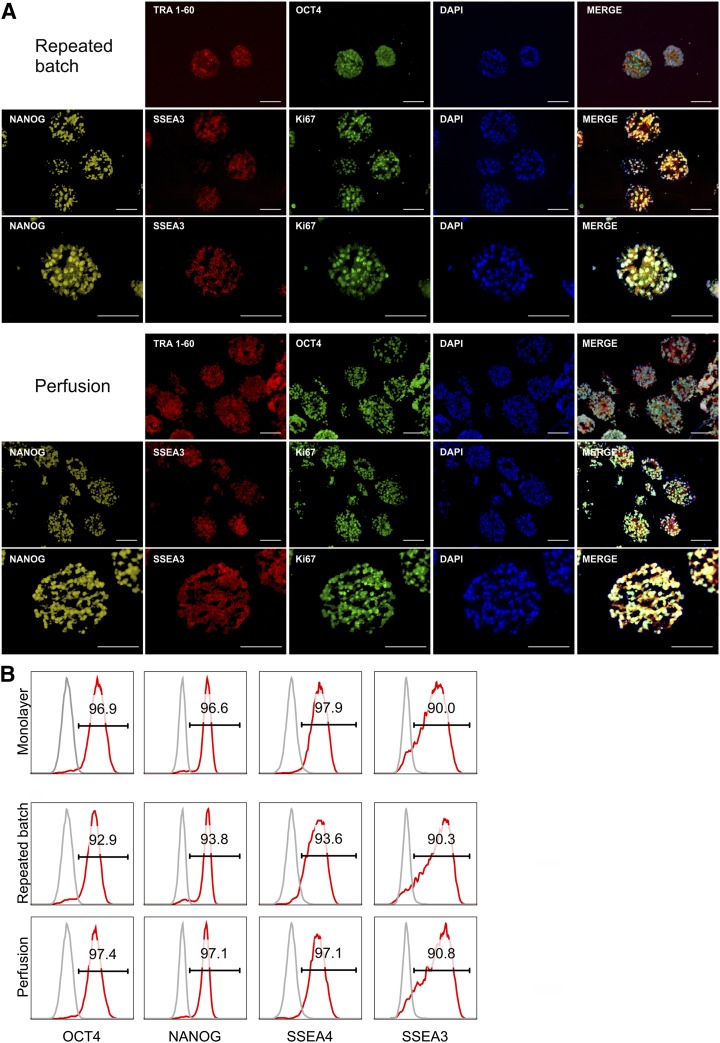
Figure 4.
Pluripotency marker expression of hCBiPS2 cells after expansion with different feeding strategies. (A): Immunofluorescence of cryosections of aggregates harvested at process day 7 showed positive staining for the pluripotency-associated markers TRA1-60, SSEA3, OCT4, and NANOG as well as the proliferation-associated marker Ki-67. Respective isotype controls confirmed specificity of staining (data not shown). Scale bars = 100 µm. (B): Flow cytometry revealed that the majority of repeated batch- and perfusion-expanded cells expressed pluripotency-associated surface markers SSEA4 and SSEA3 as well as the transcription factors NANOG and OCT4 comparable to monolayer precultures (isotype control shown in gray). Abbreviation: DAPI, 4′,6-diamidino-2-phenylindole.

