
Figure 3.
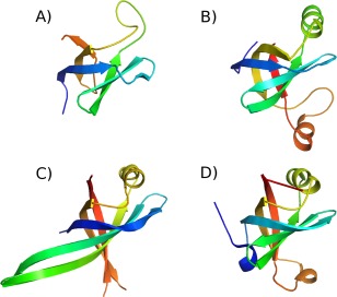
Models of the B. subtilis ComEC OB fold compared to crystal structures. Structures are shown coloured from blue (N‐terminus) to red (C‐terminus) (A) homology model of residues 72‐148. (B) covariance‐assisted fragment‐assembly model of residues 72‐167. (C) The crystal structure of Primosomal Replication Protein N Klebsiella pneumonia (PDB code 4apv; 56) D) the OB fold from subunit A of the structure of Pseudomonas aeruginosa Glutamyl‐tRNA(Gln) Amidotransferase (PDB code 4wj3; 58). [Color figure can be viewed in the online issue, which is available at wileyonlinelibrary.com.]