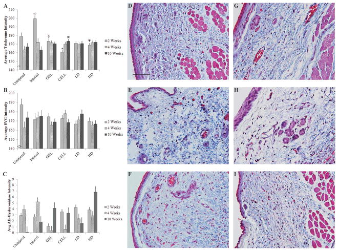
Figure 3.
Trichrome staining differed by treatment group and weeks post-injury, but there were no significant effects for EVG or Alcian Blue. Average trichrome (A), EVG (B) and Alcian Blue with hyaluronidase digestion (AB-Hyaluronidase; C) staining intensities are included for two control groups (Uninjured and Injured) and four treatment groups (GEL, CELL, LD and HD). Data are shown as mean ± standard error. *P < 0.05 compared with all other groups at 2 weeks following treatment; ∞P < 0.05 compared with Uninjured, LD at 2 weeks following treatment; ¥P < 0.05 compared with Uninjured, Injured at 2 weeks following treatment; §P < 0.05 compared with Injured at 2 weeks following treatment;
 P < 0.05 compared with Injured at 10 weeks following treatment. Representative histological images of trichrome-stained Uninjured (D), Injured (E), CELL (F), GEL (G), LD (H) and HD (I) vocal fold are shown 10 weeks following treatment (40× magnification; scale bar = 100 μm).

