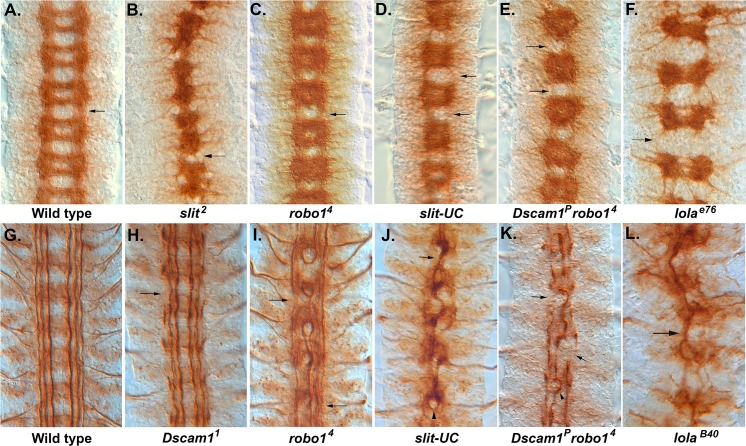
Fig 1. Longitudinal axon guidance in Dscam1, robo1, and slit-UC mutants.
Embryos stained with monoclonal antibody (mAb) BP102 (A–D) to reveal the central nervous system (CNS) axon scaffold and anti-Fas2 (1D4) to reveal the longitudinal tracts (E–L). (A) Stage 16 wild-type embryo with an intersegmental longitudinal tract marked by an arrow. (B) slit2 homozygous embryo displaying the characteristic collapse of the axon scaffold onto the CNS midline. (C) robo14 homozygous embryo showing the characteristic pattern of thickened commissures and reduced longitudinals (arrow). BP102 staining in the longitudinal tracts is reduced. (D) Stage 16 uncleavable slit mutant displaying reduced or absent longitudinals (arrows) and variability in the degree of axon collapse towards the midline in each segment, e.g., a gap between the commissures is visible in the lowest segment but not in any other segment. (E) Dscam1P robo4 mutant in which several longitudinal tracts are absent (arrows). The nerve cord has not condensed as much as wild type or the robo mutant, so less segments are visible in the field of view. The individual segments are more variable than the robo mutant. (F) lolae76, a strong allele in which the longitudinal connectives (arrow) are almost completely absent. There is also a reduction in the thickness of the commissures. (G) Stage 17 wild-type embryo with longitudinal tracts running parallel to the CNS midline (center of image). (H) Dscam11 embryo with breaks to the outermost fascicle (arrow) and an overall waviness to the longitudinal fascicles. (I) robo14 mutant in which the innermost fascicles frequently merge and form circles around the midline. The medial and lateral fascicles are present (not always in the focal plane) and largely intact but display thinning and occasional breaks and defasciculation (arrows). (J) slit-UC mutant in which many of the longitudinal axons have fused to form a large fascicle that meanders along the midline (arrow). Remnants of the outermost fascicle are present but slightly out of focus, reflecting an overall degree of disorganization to the nerve cord. The occasional robo-like circle is visible (arrowhead). (K) Dscam1P robo4 homozygous embryo displaying disruption to all the longitudinal axon tracts. A large, thick fascicle follows the midline, occasionally forming circles (arrowhead). The outermost fascicle is still present but is thicker than normal, so it has likely fused with the intermediate fascicle. There are frequent breaks, and sometimes the fascicle is completely absent (arrows). (L) lolaB40 strong allele showing a large fascicle wandering back and forth across the midline (arrow). The underlying data are shown in S1 Data.

