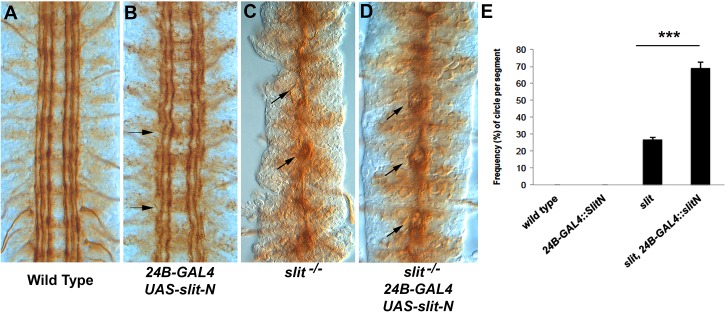
Fig 8. Partial suppression of the slit CNS phenotype by slit-N expression.
The longitudinal tracts of stage 17 embryos were stained with anti-Fas2. (A) Stage 17 wild-type control embryo. (B) Expression of slit-N lateral to the CNS using the 24B muscle-specific promoter. Some waviness of the tracts and occasional breaks (arrows) are seen, suggesting that the ectopic slit-N is disrupting normal guidance. (C) A slit mutant displaying the characteristic collapse of the tracts onto the CNS midline. An occasional separation of the fascicles, which appears as a small circle, can be seen (arrow), as opposed to a failure of motor neuron bundles to leave the CNS (arrowhead). The motor neurons are easily distinguished by their position and the absence of the motor nerve root in that segment and were not counted in analysis of phenotypes. (D) Expression of slit-N in a slit mutant induces frequent separation of the fascicles at the CNS midline, appearing as small circles (arrows). These circles are slightly larger and more frequent than those seen in the slit mutant alone. (E) Quantification of the number of circles visible in the abdominal and thoracic segments expressed as percentages for the genotypes indicated (S5 Data). We have never observed the circles in wild type or in embryos expressing slit-N by 24B-GAL4. A statistically significant difference between slit and slit with lateral expression of slit-N was determined using a Tukey HSD within a one-way ANOVA, ***p < 0.001. The underlying data are shown in S5 Data.

