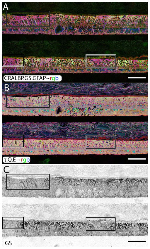
Figure 11.
A two regions of human RP retina, stacked, probed and registered in serial sections for CRALBP, GS, GFAP in A, taurine, glutamine and glutamate in B. The isolated GS channel is shown in C. All three panels demonstrate inhomogeneous levels of GS expression occurring in human RP in response to retinal degeneration and subsequent retinal remodeling. Before all photoreceptors die off, GS increases in concentration only to disappear in regions where Müller cells are engaging in seal formation. Scale = 200 μm.

