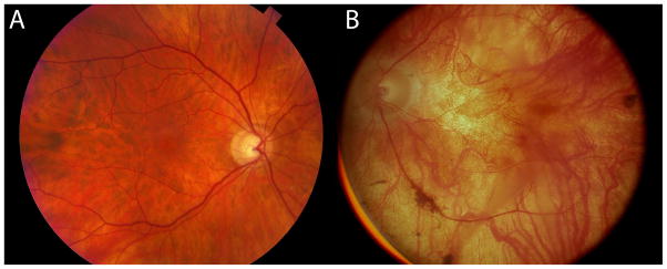
Figure 2.
Fundoscopic images of a 67 year old normal human retina (HS1) in A and a fundoscopic image from an RP donor (RP1), then aged 58 when imaged in 1991 in B, 20 years prior to death. The fundoscopic image of RP1 demonstrates a pale fundus, optic nerve atrophy, and vessel attenuation. This retina provided the tissue for sample RP1.

