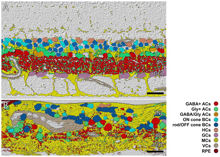
Figure 9.
ISODATA clustering of retinal cell classes. A. In normal primate retina lamination is precise (outer plexiform layer, OPL; inner nuclear layer, INL; inner plexiform layer IPL, ganglion cell layer GCL, optif fiber layer, OFL, inner limiting membrane ILM). The IPL is ≈ 40 μm thick B. Sample RP3.(republished from Jones et. al. 2003) showing remodeling clearly disrupting normal lamination of the retina. The IPL and GCL are essentially gone. Scale = 40 μm.

