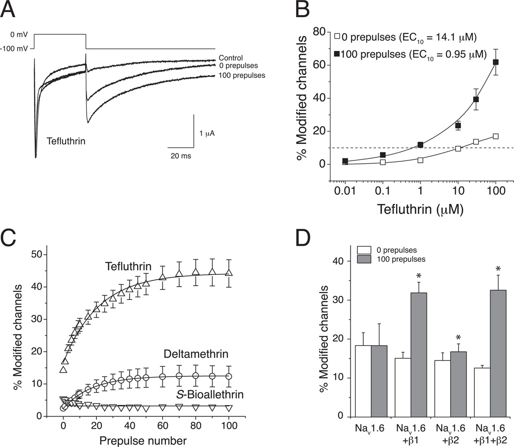
Fig. 2.
Resting and use-dependent modification of rat Nav1.6 sodium channels expressed in Xenopus oocytes by pyrethroids (redrawn from data in Tan and Soderlund, 2010; Tan and Soderlund, 2011b). (A) Representative traces recorded from an oocyte expressing rat Nav1.6+β1+β2 sodium channels exposed to 100 µM tefluthrin. The control trace was recorded prior to insecticide exposure. Following equilibration with tefluthrin traces were recorded before or after the application of a high-frequency train of 100 depolarizing prepulses (5 ms, 66.7 Hz). (B) Concentration dependence of resting (0 prepulses) and use-dependent (100 prepulses) modification of rat Nav1.6+β1+β2 sodium channels by tefluthrin. Values are means ± SE of 5 separate experiments. The dashed line indicates 10% channel modification. (C) Effect of repeated depolarizing prepulses on the extent of modification of rat Nav1.6+β1+β2 sodium channels by S-bioallethrin, tefluthrin and deltamethrin. Values are means ± SE of 4 (S-bioallethrin), 15 (tefluthrin) or 12 (deltamethrin) separate experiments with different oocytes. (D) Comparison of the extent of resting (after 0 prepulses) and maximal use-dependent (after 100 prepulses) modification of rat Nav1.6, Nav1.6+β1, Nav1.6+β2, and Nav1.6+β1+β2 by tefluthrin. Values for use-dependent modification marked with asterisks were significantly different from values for the resting modification of the same channel (paired t-tests, P < 0.05).

