Figure 1.
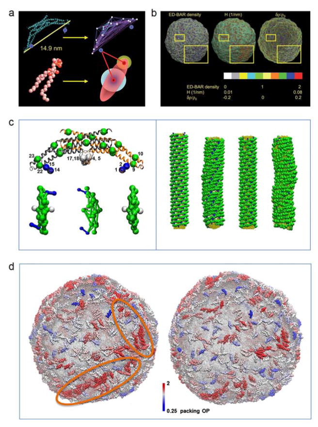
a) Schematics of heteroENM CG model of N-BAR domain (top) and HAS CG lipid model (bottom) as employed in (Ayton et al., 2010) for liposome simulations. The CG model of the N-BAR domain is derived using the ED-CG approach (Zhang et al., 2008) from atomistic simulations. Two sites are introduced to represent each amphipathic helices (highlighted by blue spheres). In the membrane the lipids are modeled as Gay-Berne ellipsoid of revolution, while the additional headgroup sites interact with all CG sites of N-BAR domain. b) The panel demonstrates the correlation between N-BAR density, mean curvature, H, and relative membrane density change, δρ/ρ0, for a selected snapshot of liposome simulations from (Ayton et al., 2010). The yellow squares point to the areas of the membrane where this correlation is most evident. c) The left side shows the schematic representation of ED-CG model of endophilin N-BAR (top) and top view of zigzag N-BAR, triad N-BAR, and BAR domain models, respectively. The zigzag and triad models correspond to different amphipathic helices orientations, while BAR lacks the amphipathic helices. The amphipathic helices are highlighted in blue, and the insert helices in white. The right side shows initial and final snapshots of CG simulations of N-BAR coated tubes. In the case of zigzag orientation (the two most left snapshots) the protein lattice remains more organized, while triad orientation (the two most right snapshots) destabilizes the lattice. d) Representative snapshots of N-BAR-coated (left) and BAR-coated (absent the amphipathic helices) (right) liposome simulations. The highlighted region shows the string-like organization of N-BAR domains that is not observed for BAR domain. Panels a) and b) are adapted from (Ayton et al., 2010) with permission from The Royal Society of Chemistry. Panels c) and d) are adapted from (Cui et al., 2013), © 2013 by the Biophysical Society.

