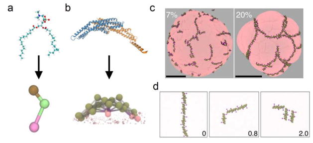
Figure 2.
Atomistic (top) and coarse-grained (CG) (bottom) representations of a) 1,2-dilauroylsn-glycero-3-phosphocholine (DLPC) lipid and b) the endophilin N-BAR domain. c) N-BAR domains form linear assemblies and meshes on the surface of liposomes inducing bud-like instabilities. The panels illustrate the remodeling of liposomes 200–300 nm in diameter at 7% (left) and 20% (right) protein surface coverages. Scale bars, 100 nm. Adapted from (Simunovic et al., 2013a), © 2013 National Academy of Sciences, USA. d) Membrane tension affects the assembly of N-BAR domains on the membrane. Shown are snapshots from simulations at different surface tensions (indicated in bottom right corner, units: mN/m) at 4% protein surface coverage. Taken from (Simunovic and Voth, 2015), © 2015 Nature Publishing Group.

