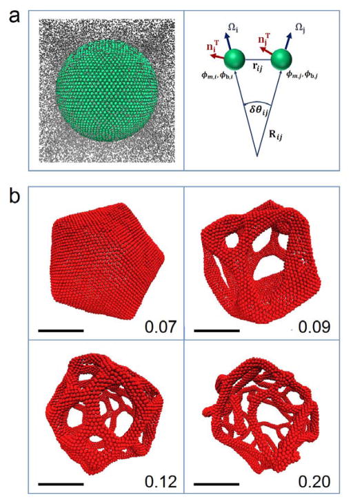
Figure 3.
a) Left: initial configuration of a mesoscopic liposome immersed in a mesoscopic solvent. Right: each membrane quasi-particle is characterized by a normal and an in-plane vector (Ω and nT, respectively), as well as lipid and protein composition fields (ϕm and ϕb, respectively). The principal component of the local curvature (1/R) along the in-plane vectors is computed on the fly based on the angles between the normal vectors (δθ) and distances between the quasi-particles. b) Final configurations of mesoscale simulations of liposomes (250 nm in diameter) fully coated by proteins for cases of various spontaneous curvatures (indicated in bottom right corner, units: 1/nm). When the spontaneous curvature is sufficiently high, the liposome undergoes a topological transition into a tubular network, with the diameter of the tubules decreasing with increasing spontaneous curvature. Scale bars, 100 nm.

