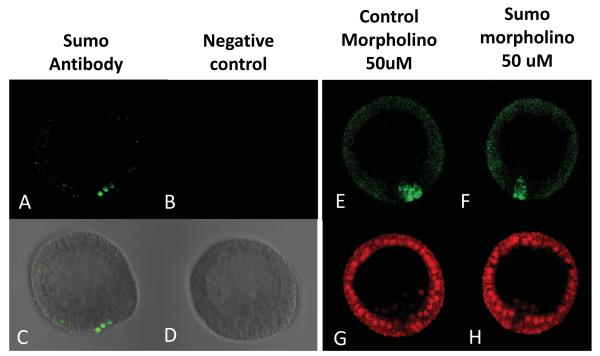
Figure 2.
Sumo protein accumulates in the small micromeres coincident with Nanos2 in blastula. Immunofluorescence using the antibody against Sumo (Abcam) was tested in blastula (A,C). Images using only the secondary antibody were used as a control (B,D), and were taken using the same microscope settings (laser intensity, pin-hole opening) at 400x magnification. Co-injection of Sp Sumo morpholino (50uM) with the GFP Nanos 2 transcript does not affect the enrichment of the GFP Nanos 2 protein in the small micromeres (F). A morpholino against Pm dysferlin was co-injected with the GFP Nanos 2 transcript as a non-relevant MO control (E). Images (E and F) were taken using the same microscope settings (laser intensity, pin-hole opening) at 400x magnification. In each case, a third transcript coding for mCherry and surrounded by Xenopus β-globin, was co-injected as a control reporter. Images (G and H) were taken using the same microscope settings (laser intensity, pin-hole opening) at 400x magnification.

