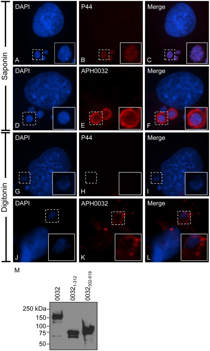
Figure 2.
APH0032 is exposed on the cytosolic face of the AVM. A. phagocytophilum infected RF/6A cells were permeabilized with either saponin (A–F), which permeabilizes both the plasma and organelle membranes to allow for antibody delivery inside organelles, or digitonin (G–L), which permeabilizes the plasma membrane but not organelle membranes. The cells were screened with APH0032 antiserum (D–F, J–L), fixed, and viewed by indirect immunofluorescence confocal microscopy. To verify that digitonin permeabilized the plasma membrane without compromising the integrity of the AVM, digitonin-treated cells were incubated with a monoclonal antibody specific for P44 (A–C, G–I), which is found on the bacterium's surface but not on the AVM. Host cell and bacterial nuclei are stained with DAPI (A,D,G,J). Bound primary antibodies were detected by Alexa Fluor 594-conjugated goat anti-rabbit IgG (B,E,H,K) and confocal microscopy. Merged images are presented in panels (C,F,I,L). Corner inset images are enlarged views of the regions denoted by hatched line boxes. (M) APH0032 antiserum recognizes both the non-repeat and tandem repeat portions of APH0032. Whole cell lysates of HeLa cells transfected to express GFP-tagged APH0032 or portions thereof were Western blotted and screened with APH0032 antiserum. Results are representative of at least two separate experiments with similar results.

