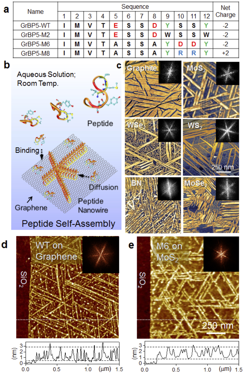
Figure 1. Atomic force microscope (AFM) images of self-assembled peptides.
(a) Sequences of the Wild Type and variant peptides. (b) A schematic showing the peptide self-organization with a series of the surface processes: binding, diffusion and self-organization. (c) AFM images of organized peptides on surfaces of bulk graphite, MoS2, WSe2, WS2, MoSe2, and hBN. All morphology shows six-fold symmetry. (d) AFM of WT on single-layer graphene. The bright lines are self-assembled peptides forming organized nanostructures. (e) AFM image of M6 on single-layer MoS2. The insets: fast Fourier transform of the AFM image exhibiting six-fold symmetry of the self-assembled peptides.

