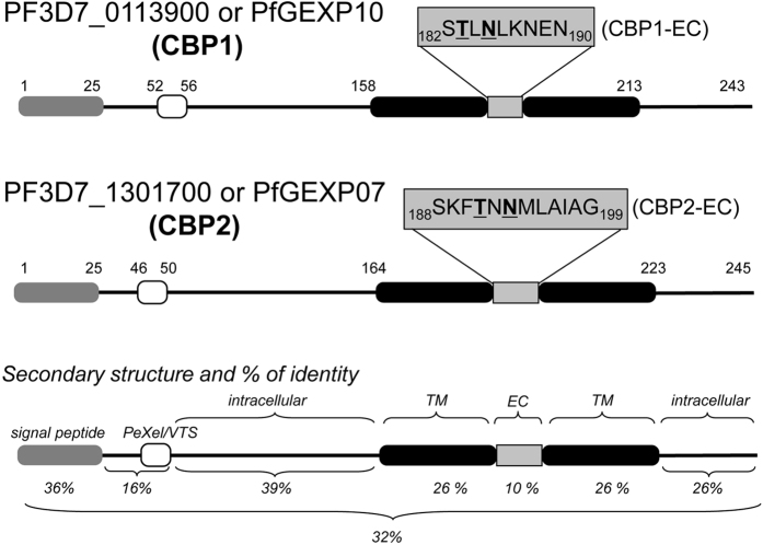
Figure 4. Schematic primary and secondary structure of CBP1 and CBP2.
Grey box: signal peptide. Empty box: Pexel/VTS motif. Black box: putative transmembrane domain. The % number indicates the sequence identity between CBP1 and CBP2 domain by domain or as a whole (lower). EC means putative “extracellular domain”.

