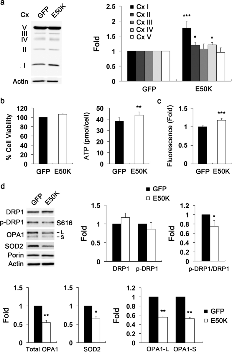
Figure 6. Overexpression of E50K alters OXPHOS Cx system, increases ROS production, and triggers mitochondrial fission and OPA1 loss in RGCs in vitro.
Primary RGCs were transduced with AAV2-GFP or AAV2-OPTNE50K-GFP for 2 days in vitro. (a) Immunoblot analysis of OXPHOS Cx protein in cultured RGCs overexpressing GFP (control) or E50K mutant. (b) Analyses of cell viability using the MTT assay and cellular ATP production using a luciferase-based assay in cultured RGCs overexpressing GFP (control) or E50K mutant. (c) ROS measurement in cultured RGCs overexpressing GFP (control) or E50K mutant. (d) Immunoblot analyses of DRP1, phospho-DRP1 (S616), OPA1, SOD2 and porin protein in cultured RGCs overexpressing GFP (control) or E50K mutant. For each determination, the actin level in the control was normalized to a value of 1.0. Data are shown as the mean ± S.D. (n = 3 independent experiments). *P < 0.05; **P < 0.01; ***P < 0.001 compared with the control group. Full-length blots are presented in Supplementary Figure 8. CypD, cyclophilin D; Cx, complex; DRP1, dynamin-related protein 1; GFP, green fluorescent protein; L, large form; S, small form; OPA1, optic atrophy type 1; OPTN, optineurin; RGC, retinal ganglion cell; ROS, reactive oxygen species; S616, serine 616; SOD2, superoxide dismutase 2.

