. 2016 Jul 31;49(7):376–381. doi: 10.5483/BMBRep.2016.49.7.246

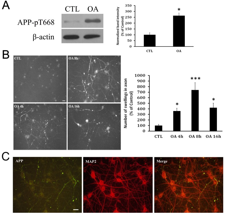
Fig. 1. Increased level of phosphorylated APP in OA-treated neuron cultures. (A) Western blot analysis with phosphorylated-amyloid precursor protein (APP) antibody shows that APP phosphorylation meaningfully increased in okadaic acid-treated neurons compared with control neurons. (B) Temporal changes in level of phosphorylated APP in the neurite swellings. Control neurons show reduced immunoreactivity for APP. APP immunoreactivity was increased in the neurite swellings within 4 hr post treatment of OA; APP immunoreactivity was found to be accumulated in cell bodies at 16 hr post-treatment. Scale bar represents 20 µm. (C) APP (+) swellings in neurites does not colocalize with MAP2 (+) dendrites. Scale bar represents 20 µm.

Fig. 1.