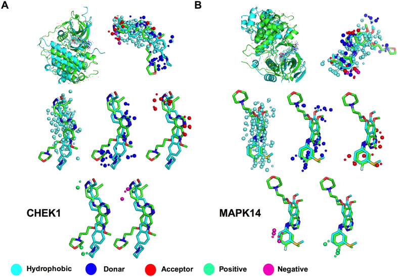
Figure 2. Binding site structure similarity between query protein and off-targets CHEK1 and MAPK14.
The superimposed structures of query and off-target proteins in complex with respective ligands are shown (A,B; top, left panel). The similarity of binding pockets in query (green) and off-targets (cyan) are shown (A,B; top, right panel). The hydrophobic, donor, acceptor, negative and positive binding site probes are shown separately and are represented by cyan, blue, red, magenta and green colored spheres, respectively (A,B; middle and lower panel). Large spheres represent the query binding probe while smaller ones represent the off-targets binding probe.

