
Figure 8. TRPV1 and the CB1 receptor are engaged in PSN.
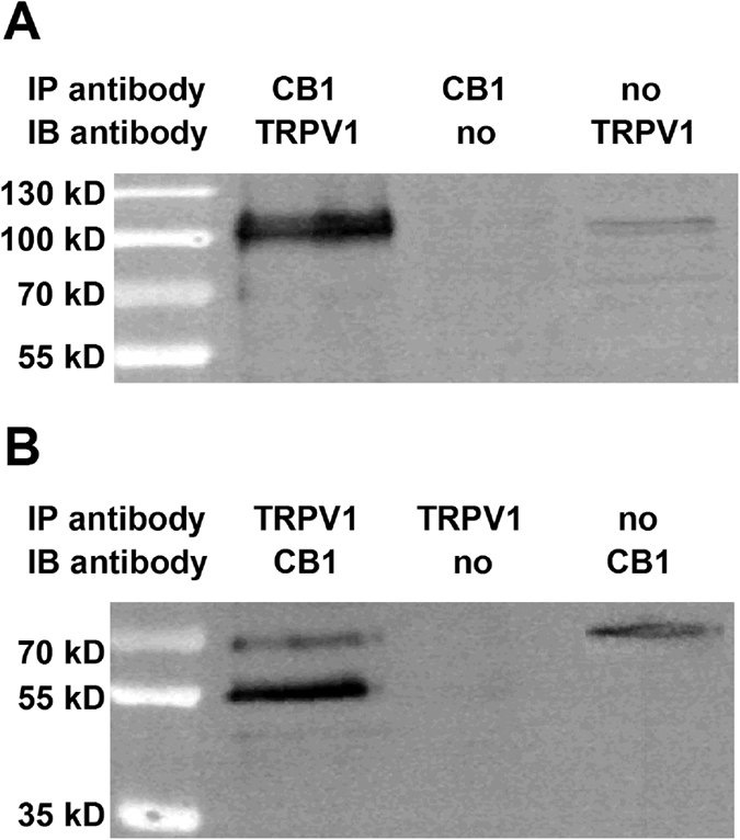
Immunoblots showing TRPV1/CB1 receptor heteromers in the rat DRG. Whole cell lysates were prepared from DRG and immunoprecipitation assays using anti-CB1 (A) and anti-TRPV1 (B) antibodies were carried out as detailed in Materials and Methods. The resulting immune complexes were analysed by immunoblotting using anti-TRPV1 (A) and anti-CB1 (B) antibodies. Immunoprecipitating antibodies and immunoblotting antibodies were omitted from positive and negative controls, respectively. TRPV1 was detected as a ~100 kDa band, while the CB1 receptor was detected as a doublet with molecular weight ~55 and ~63 kDa in the immunoprecipitated samples.