Figure 1.

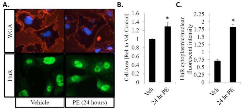
HuR nucleo-cytoplasmic shuttling correlates with an increase in cell size/hypertrophic growth in neonatal rat ventricular myocytes (NRVMs). Following a 24-hour exposure to PE (10 μM), NRVMs show a significant increase in cell area as indicated by WGA staining (A, top panel) and cytoplasmic translocation of HuR as determined by HuR IHC (A, bottom panel). (B) Cell surface area was quantitatively determined using NIH Image J, and is expressed as fold-increase in area compared to vehicle control treated cells. (C) HuR translocation was quantified as the ratio of cytoplasmic to nuclear fluorescent intensity. N ≥ 4 for each group (each N represents the average measurement of 10 cells per well). *P ≤ 0.05 vs. Veh control.
