
Fig. 1.
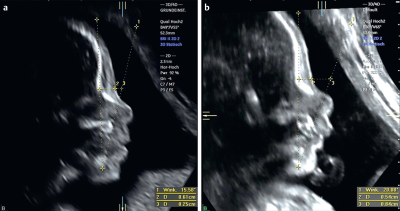
Fetal profiles obtained from facial volumes of a trisomy 21 fetus a and an euploid fetus b at 22 weeks of gestation. Left panel corresponds to the coronal view with the reference dot placed between the nostrils. Sagittal plane showing 2 lines tracing the mandible, the nasion and the anteriormost border of the maxilla, respectively. The resulting novel MMN angle is significantly more acute in a Down syndrome fetus. In addition, the PFSR is markedly decreased (panel a) in this fetus compared to controls (panel b).