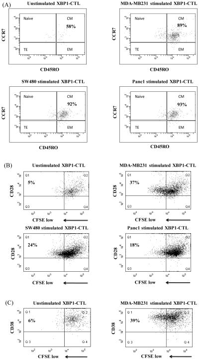
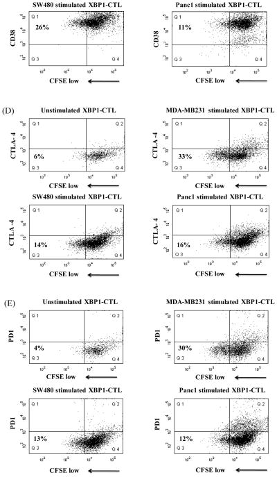
Figure 5. Lenalidomide increased specific XBP1-CTL proliferation to breast cancer, colon cancer or pancreatic cancer cells.
Specific cell proliferation was examined by flow cytometry after stimulating CFSE-labeled XBP1-CTL with tumor cells for 6 days. The proliferating CD3+CD8+T cell population was measured as the percent decrease in CFSE expression. Enhanced cell proliferation of lenalidomide treated (5 μm, 4 days) XBP1-CTL was examined in response to irradiated respective tumor cells. Background proliferation was determined using CFSE-labeled lenalidomide treated XBP1-CTL cultured in media alone. (A) The major proliferative response of XBP1-CTL after lenalidomide treatment was detected within the central memory (CCR7+CD45RO+) CTL subset against breast cancer (MDA-MB231), colon cancer (SW480) or pancreatic cancer (Panc1) cells. Phenotypic characterization demonstrates that specific cellular proliferation (Q1 gated) to HLA-A2+ breast cancer (MDA-MB231), colon cancer (SW480) or pancreatic cancer (Panc1) cells express (B) CD28 and (C) CD38, but neither (D) CTLA-4 nor (E) PD-1.


