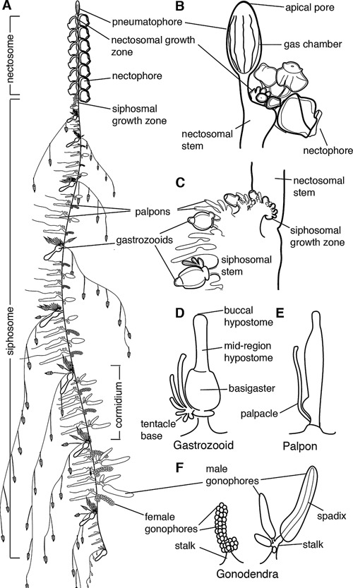
Figure 1.

Schematic of Nanomia bijuga colony. Adapted from http://commons.wikimedia.org/wiki/File:Nanomia_bijuga_whole_animal_and_growth_zones.svg, which was drawn by Freya Goetz. (A)–(C) are oriented with anterior to the top and ventral to the left. (D)–(F) are oriented with anterior to the left and ventral up. (A) Overview of the mature colony. All zooids are produced from two growth zones, one at the anterior end of the siphosome, immediately posterior to the pneumatophore, and one at the anterior end of the siphosome, immediately posterior to the nectosome. Zooids are organized on the siphosome into reiterated units known as cormidia. (B) The pneumatophore and nectosomal growth zone at the anterior of the colony, showing forming nectophores. (C) The siphosomal growth zone and budding cormidia, with gastrozooids representing cormidial boundaries. (D) Gastrozooid, connected at the base to the siphosomal stem, showing labelled regions of the mouth (buccal hypostome), the mid‐region of the hypostome, and the basigaster. (E) Palpon, connected to the stem by its peduncle. (F) Female and male gonodendra. Multiple individual gonophores are borne on stalks which connect to the siphosomal stem.
