Figure 6.

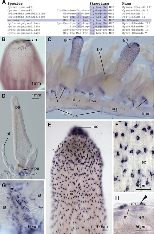
Nerve cells. In situ nb‐rfamide hybridization. (A) Table showing amino acid structures of selected cnidarian neuropeptides, modified from Grimmelikhuijzen (2004). Amino acids shared with Nb‐RFamide are shaded. (B) The pneumatophore with the anterior facing upward, showing apical pore (ap). nb‐rfamide positive cells are scattered most densely in a ring posterior to the apical pore, and less densely toward the mid region of the pneumatophore. (C) The cormidia of the siphosome, anterior to the left, with gastrozooids (ga) and palpons (pa) ventrally attached to the stem (st), and the giant axon (ax) on the dorsal side. Bracteal scars (bsc) are visible. nb‐rfamide expression can be observed in rings (r) around palpon and gastrozooid peduncles above their point of attachment on the siphosomal stems. (D) Palpon showing rings of nb‐rfamide positive cells at the base of the peduncle (pe) and at the base of the palpacle (pl). No nb‐rfamide positive cells are observed in the palpon body or palpacle. (E) Gastrozooid hypostome showing ectodermal nb‐rfamide positive cells with higher densities near the mouth (mo) and lower density in the mid‐region (mr) of the hypostome. (F) Enlarged image of ectodermal (ec) neuronal cells in the hypostome of the gastrozooid. (G) Whole mount of the siphosomal stem showing transverse collars of nb‐rfamide positive cells. (H) Whole mount of ectoderm, mesoglea (m), and endoderm (en) in the hypostome of the gastrozooid. nb‐rfamide signal is absent in the endoderm. Morphology of ectodermal nb‐rfamide positive cell (arrow) suggests sensory function.
