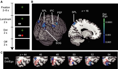
Figure 3.

Designs and results of Experiment 2 with a small landmark. (A) A large rectangular frame in Experiment 1 was reduced to the size of the dot (a small rectangle – Landmark) in Experiment 2. The conditions were otherwise the same as in Experiment 1 (Fig. 1A). (B) Regions with significant adaptation (voxel‐level P < 0.005, uncorrected; cluster‐level P < 0.05, uncorrected) as a result of repeated presentation of the dot, in terms of the egocentric (eye‐ and head‐centered) coordinates (Dot/Ego). Adaptation in the right SPL was significant after correction for the FWER (cluster‐level P = 0.035, FWER‐corrected). FEF, frontal eye field; IPC, inferior parietal cortex. (C) The right SPL region superimposed on sequential axial slices. The precuneus region found in Experiment 1 is shown by red lines. Note that there is little overlap between the two.
