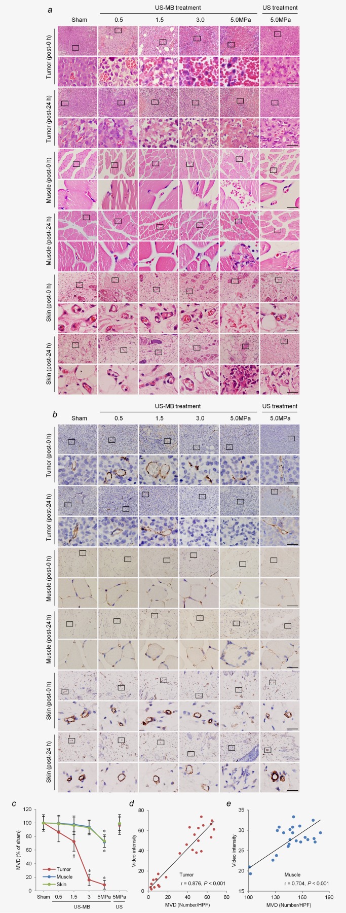
Figure 3.

Effects of LIUS‐MB treatment on microvessels in the tumor, muscle, and skin, as well as their relationships with corresponding blood perfusion: results for various pressures. Hematoxylin‐eosin (a) and immunohistochemical staining for the endothelial marker CD31 (b) in the tumor, muscle, and skin. (c) Quantitative analysis of the MVD at 24 hr after treatment. Correlations between VI data obtained by CEU imaging and the MVD at 24 hr after treatment in the tumor (d) and muscle (e). # p < 0.05, *p < 0.01 vs. the respective sham groups. MVD, microvessel density; HPF, high power field; VI, video intensity; CEU, contrast‐enhanced ultrasound. Scale bar = 20 μm. [Color figure can be viewed in the online issue, which is available at wileyonlinelibrary.com.]
