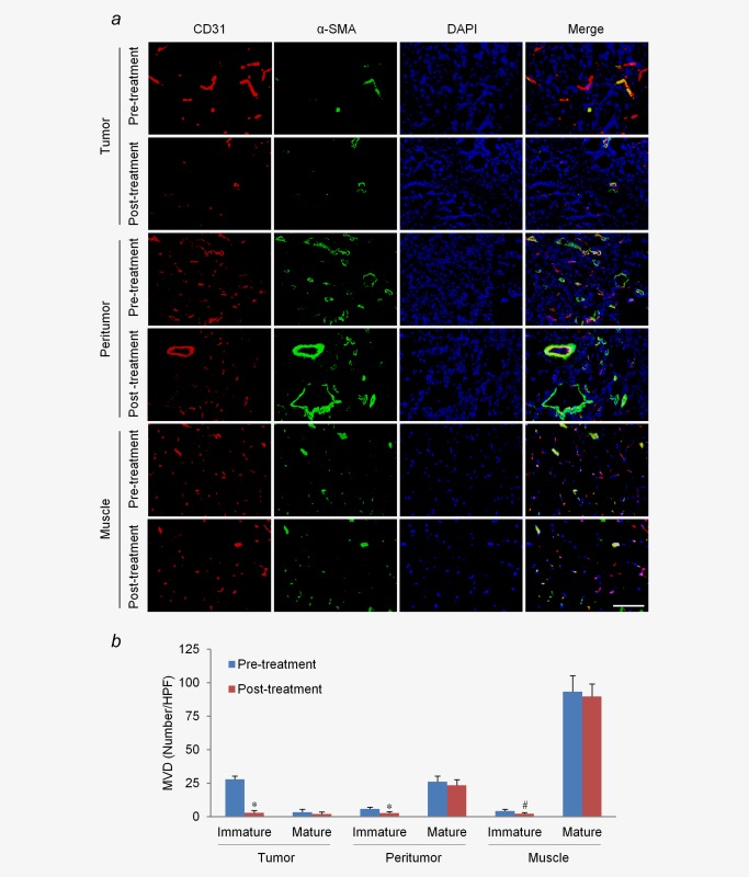
Figure 4.

Effects of LIUS‐MB treatment with 3.0 MPa on vessels of different maturity. (a) Confocal immunofluorescence of vessels in the tumor, peritumor tissue, and muscle for CD31 (red) and α‐SMA (green). (b) Quantitative analysis of immature and mature microvessels. # p < 0.05, *p < 0.01 vs. their corresponding baselines (pretreatment). SMA, smooth muscle actin. Scale bar = 100 μm. [Color figure can be viewed in the online issue, which is available at wileyonlinelibrary.com.]
