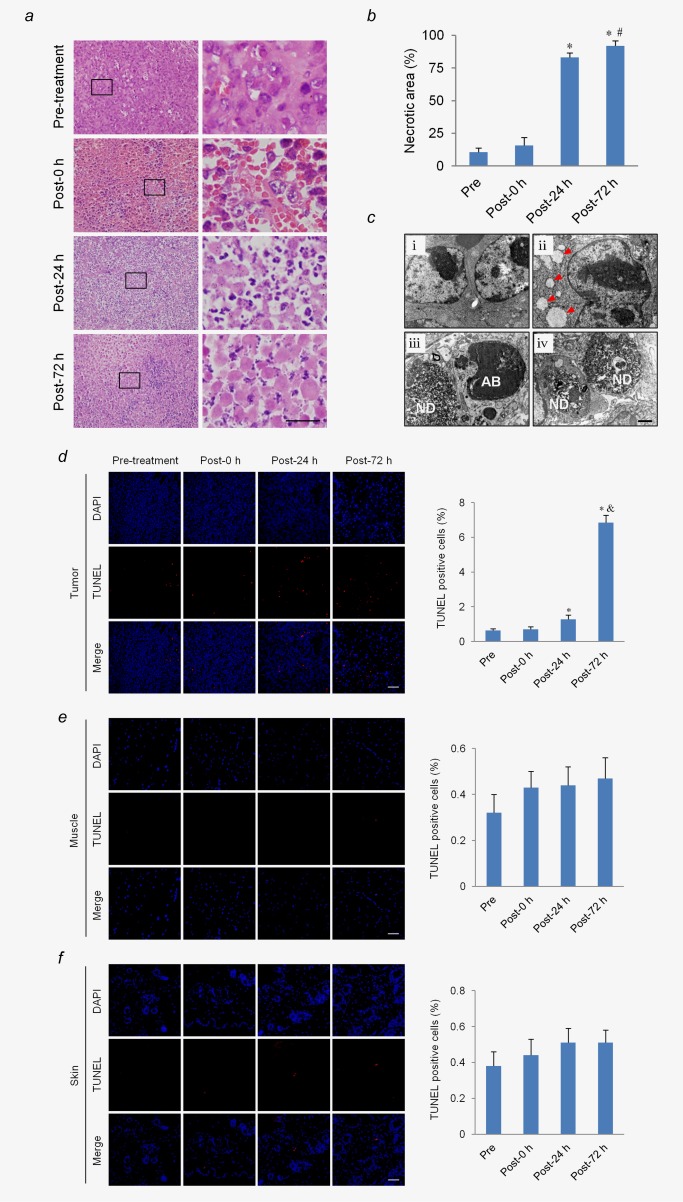
Figure 5.

LIUS‐MB treatment promotes cell necrosis and apoptosis in tumor, but not in muscle or skin. Hematoxylin‐eosin staining (a) and quantification of the necrosis area (b) in the tumor. Scale bar = 20 μm. (c) Representative transmission electronic micrographs of tumor cells before treatment (i), as well as 0 (ii), 24 (iii), and 72 hr (iv) after treatment. Ultrastructural pathological changes were observed after treatment, including mitochondria swelling, vacuolation (ii; arrows), nuclear debris (iii, iv; ND) and apoptotic bodies (iii; AB). Scale bar = 0.5 μm. TUNEL staining and quantification of the apoptotic cells in the tumor (d), muscle (e), and skin (f). Scale bar = 50 μm. *p < 0.01 vs. pre‐treatment. # p < 0.05, & p < 0.01, vs. post‐24 hr. [Color figure can be viewed in the online issue, which is available at wileyonlinelibrary.com.]
