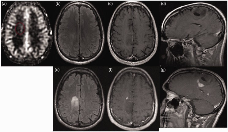
Figure 2.
A 34-year-old man with history of treated WHO Grade II glioma nine years earlier presented with seizures. (a) ASL imaging demonstrates subtle hyperperfusion while (b) FLAIR and ((c) and (d)) T1W-CE images are unrevealing. Two months later tumor progression is readily evident on (e) FLAIR and ((f) and (g)) T1W-CE images (ASL images were not obtained at that time). Biopsy revealed hypercellular infiltrating WHO Grade IV glioblastoma positive for Ki-67 labeling, IDH1 mutation and p53 immunoreactivity. Even subtle changes on ASL, 34 ml/100 g/min in this case, may represent sentinel evidence of progression, particularly when the rCBF ratio is elevated, which was 1.9 in this case.
WHO: World Health Organization; ASL: arterial spin labeling; FLAIR: fluid-attenuated inversion recovery; T1W-CE: contrast-enhanced T1-weighted; IDH1: isocitrate dehydrogenase 1; rCBF: relative CBF.

