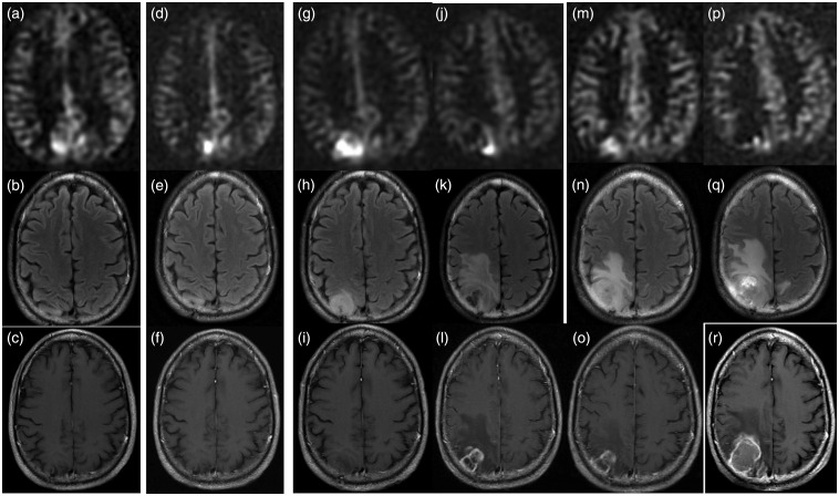
Figure 3.
A 50-year-old male with treated WHO Grade II oligodendroglioma was treated with surgery, chemotherapy and radiation eight years previously. Surveillance scan demonstrates (a) ASL-positive nodule in the right precuneus, while the (b) FLAIR and (c) T1W-CE scans are unrevealing. The (h) FLAIR and (i) T1W-CE images became concerning seven months after the onset of hyperperfusion, and it was at this point adjuvant chemotherapy and radiation were re-initiated. ASL signal appears to scale roughly with the growth of solidly enhancing tumor, while the treatment-related change anterior to the tumor evident at 11 months demonstrates no hyperperfusion ((j)–(l)). By the time the patient underwent re-resection the lesion was quite necrotic and CBF was only 22 ml/100 g/min ((p)–(r)). Histopathology demonstrated WHO Grade IV glioblastoma with oligodendroglial components, densely cellular tumor with pseudopalisading necrosis and no treatment-related necrosis. Genetics demonstrated 1p and 19q deletions and IDH1 mutation.
WHO: World Health Organization; ASL: arterial spin labeling; FLAIR: fluid-attenuated inversion recovery; T1W-CE: contrast-enhanced T1-weighted; IDH1: isocitrate dehydrogenase 1.

