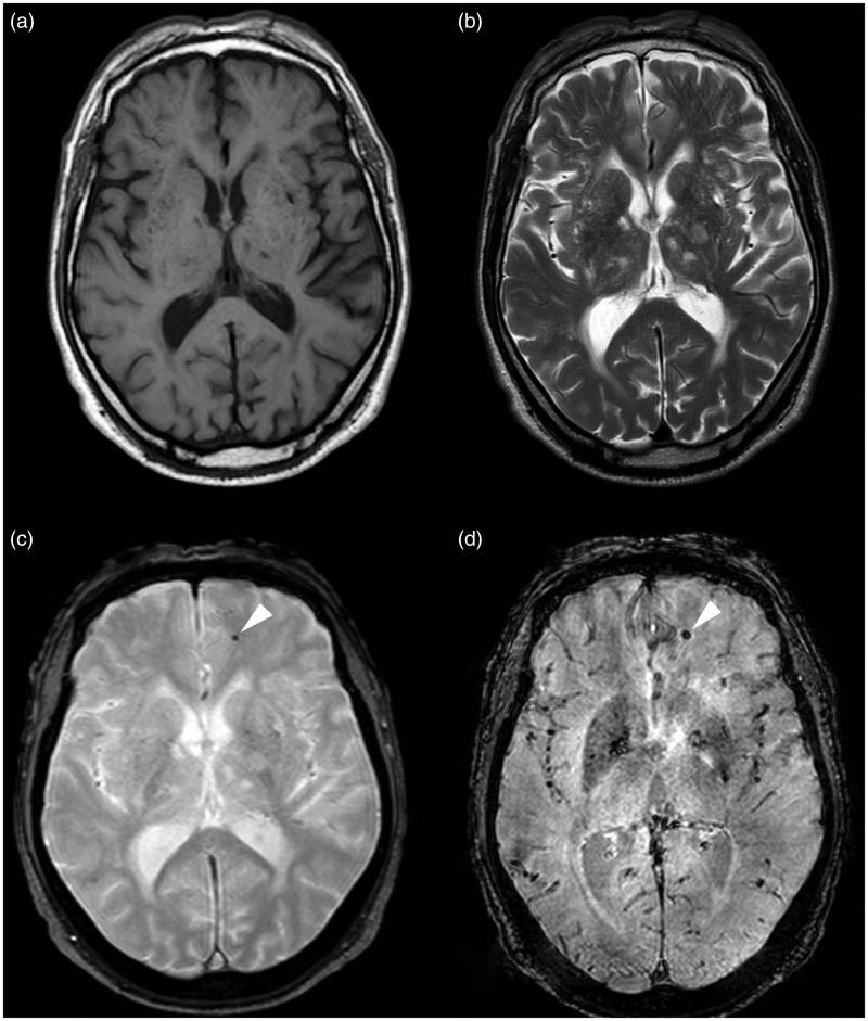
Figure 4.
MR images of Type IV lesion. The Type IV lesion (arrowhead) is not seen on (a) T1-weighted and (b) FSE T2-weighted images; while it is seen only on the (c) T2*-weighted GRE and (d) SWI images as a hypo-intense punctate area.
FSE: fast-spin echo; GRE: gradient echo images; MR: magnetic resonance; SWI: susceptibility-weighted imaging

