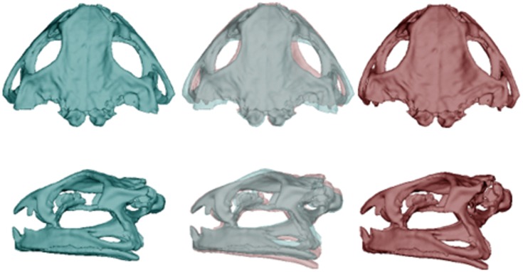
Fig 1. Differences in morphology of the skull between populations of cane toads, based on analyses of 52 specimens.
Dorsal and lateral views depict mean skull morphology of toads from long-colonised areas (left, blue) and those from invasion-front populations (right, red). The central images overlay the ones on either side to reveal points of divergence, in this case reflecting the transformation from a low (-0.06) to high (0.06) PC1 score.

