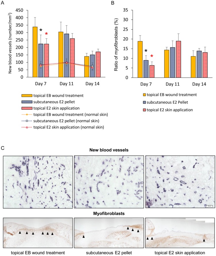
Fig 4. New blood vessels and wound contraction.
(A) The number of new blood vessels per mm2 and (B) ratio of myofibroblasts (%) are shown in box graphs. Values are expressed as the mean ± SD, n = 5–6 for each group, ANOVA, Tukey-Kramer *p<0.05 (in black): the topical EB wound treatment group versus the subcutaneous E2 pellet group, *p<0.05 (in red): the topical EB wound treatment group versus the topical E2 skin application group. (C) New blood vessels stained with an anti-CD31 antibody (bars, 50 μm) and myofibroblasts (arrows) stained with an anti-α-SMA antibody (bars, 200 μm) were observed in granulation tissue on day 7.

