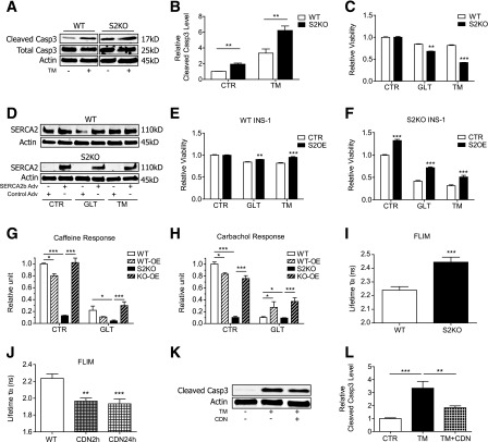
Figure 8.
SERCA2 KO β-cells manifest impaired Ca2+ storage and increased susceptibility to stress-induced death. A and B: WT and S2KO INS-1 cells were treated with or without 10 μmol/L TM for 24 h. Immunoblot analysis was performed using antibodies against cleaved caspase-3 (Casp3), total caspase-3, and actin. Quantitative protein levels are shown graphically. C: WT and S2KO INS-1 cells were treated under control (CTR) conditions or exposed to GLT or TM for 24 h, and viability was measured using the CellTiter-Glo assay. Results were normalized to results obtained in WT cells under control conditions. D–F: SERCA2b was overexpressed (S2OE) via adenoviral (Adv) transduction in WT and S2KO INS-1 cells and then treated with GLT or TM for 24 h. D: Immunoblot analysis was performed using antibodies against SERCA2 and actin. CellTiter-Glo viability tests were performed in WT (E) and S2KO cells (F). Results were normalized to cells transduced with LacZ virus under control conditions. SERCA2b was overexpressed via adenoviral transduction in WT (WT-OE) and S2KO (KO-OE) INS-1 cells, followed by exposure to GLT for 24 h. G and H: To assess cytosolic Ca2+ levels, calcium 6 fluorescence was measured under Ca2+-free conditions. I and J: INS-1 cells were transduced with a D4ER adenovirus, and FLIM was used to measure ER Ca2+. Shown is the average donor lifetime in WT and S2KO INS-1 cells (I) or WT INS-1 cells treated with 10 μmol/L CDN1163 for 2 or 24 h (J) (n = at least 10 regions of interest per cell type or treatment). K and L: WT and S2KO INS-1 cells were treated with or without TM combined with or without 10 μmol/L CDN1163 for 24 h. Immunoblot analysis was performed using antibodies against cleaved caspase-3 and actin, and quantitative protein levels are shown graphically. Results are displayed as means ± SEM. Indicated comparisons were significantly different: *P < 0.05; **P < 0.01; ***P < 0.001.

