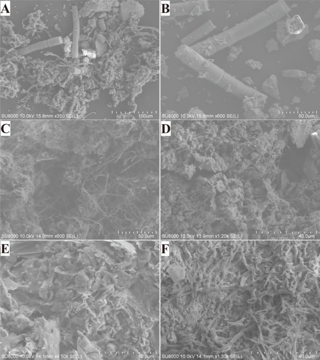
Fig 2. Scanning electron micrographs of microbial colonies from the stone monuments.
A and B show algae and the fungal hyphae forming an aggregate with the archaeological monument’s matrix in sample KH1. The images presented in C and D show Actinobacteria filaments with a diameter of 0.5–2 μm and fungal filaments with a diameter of 2–4 μm in samples LY2 and LY3. E and F show lichens growing on the surface of the stone statues and forming a foliated structure in samples QX3 and QX7.

