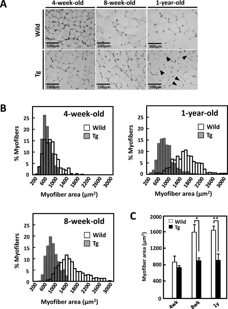
Fig 1. Time-course morphological alterations of type II fiber-rich muscles in GDE5dC471 mice.
A, Representative photographs of H&E staining in cross sections of gastrocnemius muscle, Left; 4-week-old GDE5dC471 mice (Tg) and age-matched control mice (Wild), center; 8-week-old, and right; 1-year-old. Central nuclei (arrow) appeared only in 1-year-old GDE5dC471 mice. B, Time-course alterations of fiber areas of gastrocnemius muscle of 4-week-old GDE5dC471 mice (Tg) and age-matched control mice (Wild). To visualize frequency of distribution of each myofibril, histogram images were analyzed. Both 8-week-old and 1-year-old Tg showed a leftward shift, indicating an evident increase in the percentage of small areas compared with age-matched Wild. C, Mean fiber areas of gastrocnemius muscle of 4-week-old GDE5dC471 mice (Tg) and age-matched control mice (Wild). 4-week-old: n = 4, 8-week-old: n = 3, and 1-year-old: n = 7 (Wild) and n = 10 (Tg). Data represent mean ± SD. *p<0.05, **p<0.01.

