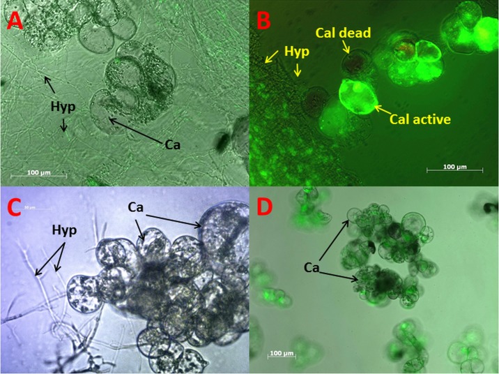
Fig 2. Fluorescence microscopy of V. vinifera callus culture and P.ch mycelium.
A: GFP labeled P.ch visible as green hyphae (Hyp), and callus cells (Cal). B: Fluorescein-diacetate stained active callus cells and dead callus cells (none fluorescent). C: Brightfield image showing hyphae growing around callus cells. D: Monoculture of callus cells with live staining (fluorescein-diacetate).

