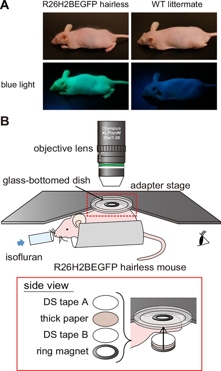
Fig 1. Our intravital imaging method using the newly established R26H2BEGFP hairless mice.
(A) Photograph of a R26H2BEGFP hairless mouse and a WT littermate. The upper and lower panels show each mouse under ordinary white light and blue light. (B) A schematic showing the intravital imaging of the dorsal skin with an upright two-photon microscope. The inset in the red rectangle shows the region indicated by the red dashed rectangle in the upper image viewed from the side. The detailed procedure is provided in the Materials and Methods and S2 Fig.

