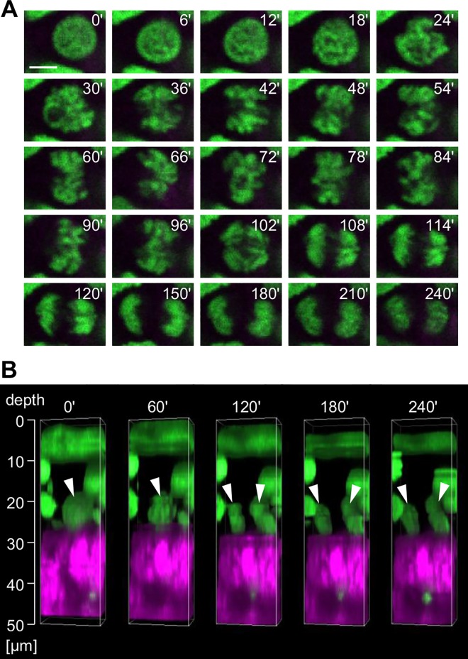
Fig 4. Four-dimensional imaging of cell division in the dorsal epidermis.
(A) Time-lapse images of the basal cells of the dorsum at a 24-μm depth from the skin surface. Images that were recorded every 6 min or 30 min are shown. These images were detected with sufficient sensitivity to determine the mitotic stage from the pattern of chromatin. It is presumed the cell is in prophase at 0–18 min, prometaphase at 24–36 min, metaphase at 42–96 min, early anaphase at 102 min, anaphase at 108–120 min, and probably telophase at 120–240 min. Scale bar = 5 μm. (B) Reconstructed three-dimensional images. The white arrowheads indicate the dividing cell. Images taken every 60 min from 0 min to 240 min in A were used for the reconstruction.

