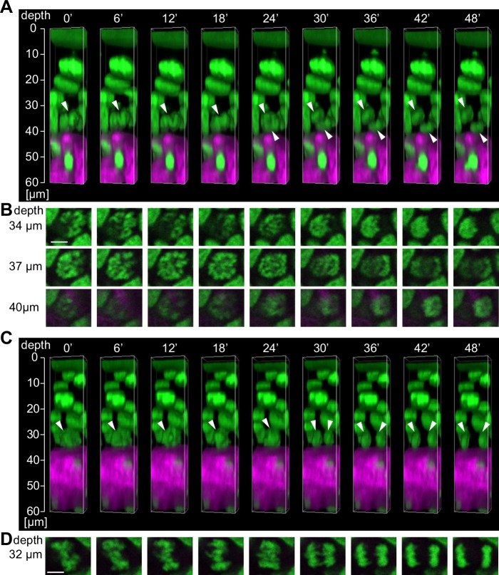
Fig 5. Four-dimensional imaging of cell division in the hind paw epidermis in a living mouse.
(A) Reconstructed three-dimensional images of the oblique division to the basement membrane from images taken every 6 min. The white arrowheads indicate a dividing cell. (B) Time-lapse images of the mitotic cells shown in A in the x-y plane at each depth. Scale bar = 5 μm. (C) Reconstructed three-dimensional images of the division parallel to the basement membrane from images taken every 6 min. The white arrowheads indicate the dividing cell. (D) Time-lapse images of the mitotic cells shown in C in the x-y plane at a depth of 32 μm from the skin surface. Scale bar = 5 μm.

