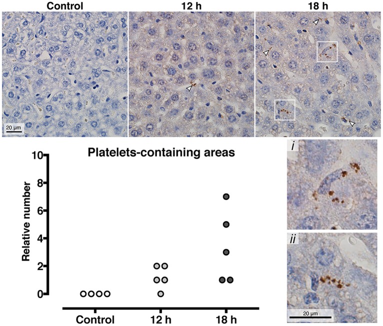
Fig 5. Platelets are progressively accumulated in the liver sinusoids at during invasive infection.
At 0, 12, and 18 hours post S. pyogenes infection the mice were sacrificed and the liver was harvested, fixed and embedded in paraffin blocks, sectioned and immunohistochemistry was performed with anti CD41 to detect platelets. Representative images are shown and quantification of the number of platelet aggregates per slide is illustrated as a column graph, where each dot is an individual mouse.

