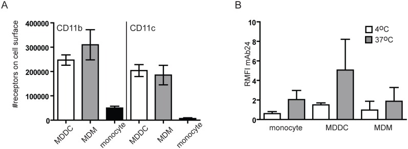
Fig 1. Expression and conformation of CD11b and CD11c.
(A) The exact amount of CD11b and CD11c on the surface of monocyte-derived dendritic cells (MDDC), monocyte-derived macrophages (MDM) and monocytes were determined using Qifikit (Dako) as described in Materials and methods. Data presented are mean +/-SD of three independent donors’ results. (B) Cells were stained with monoclonal antibody mAb24 that is specific for the high affinity conformation of CD18. Relative mean fluorescence intensity (RMFI) was calculated in each case by comparing the signal of mAb24 stained cells to isotype matched control antibody stained cells (RMFI = MFI mAb24/MFI isotype control). At 4°C RMFI values were around 1 (monocytes: 0,60+/-0,22; MDDC: 1,50+/-0,20; MDM: 0,97+/-0,91), meaning that cells do not have active β2 integrins on their surface. At 37°C all cell types bound mAb24 (RMFI for monocytes: 2,05+/-0,93; MDDC: 5,07+/-3,15; MDM: 1,87+/-1,42) showing that β2 integrins were in a conformation capable of ligand binding on their surface. Data presented are mean +/-SD of three independent donors’ results.

