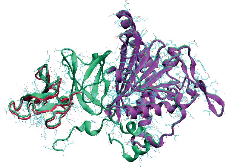
Fig 9. Substructures of 1,9a-dioxygenase (PDB id: 1WW9) that were used as an initial test of the force fields.
More extensive tests of the full complex (PDB ids: 2DE5-7) will be reported elsewhere. Left(red), the Rieske region with [2Fe-2S]. Right(purple), the region encompassing the mono-nuclear Fe structure. The [2Fe-2S] half is the structure from PDB id 3GKQ (a related 1,9a-dioxygenase from Sphingomonas, strain KA1 which uses a 4-Cys-[2Fe-2S] Ferredoxin cognate). The simulations are shown in Fig 10.

