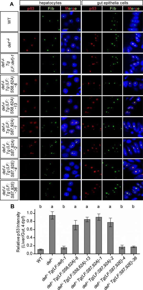
Fig 10. Simultaneous phosphorylations at S58 and S62 or at S87 and S92 are essential for Def to mediate p53 degradation in the nucleoli.
(A) Co-immunostaining of p53 (in red) and Fib (in green) in different genotypes as shown. DAPI was used to stain the nuclei (blue). Scale bar: 5 μm. (B) Statistical data showing the fold change of p53 signal intensity for hepatocytes versus intestinal epithelial cells in (A). Data are presented as means with SEM. Columns with no common letter are significantly different (p < 0.001, one-way ANOVA with Tukey’s post hoc test). Underlying data for (B) are provided in S1 Data.

