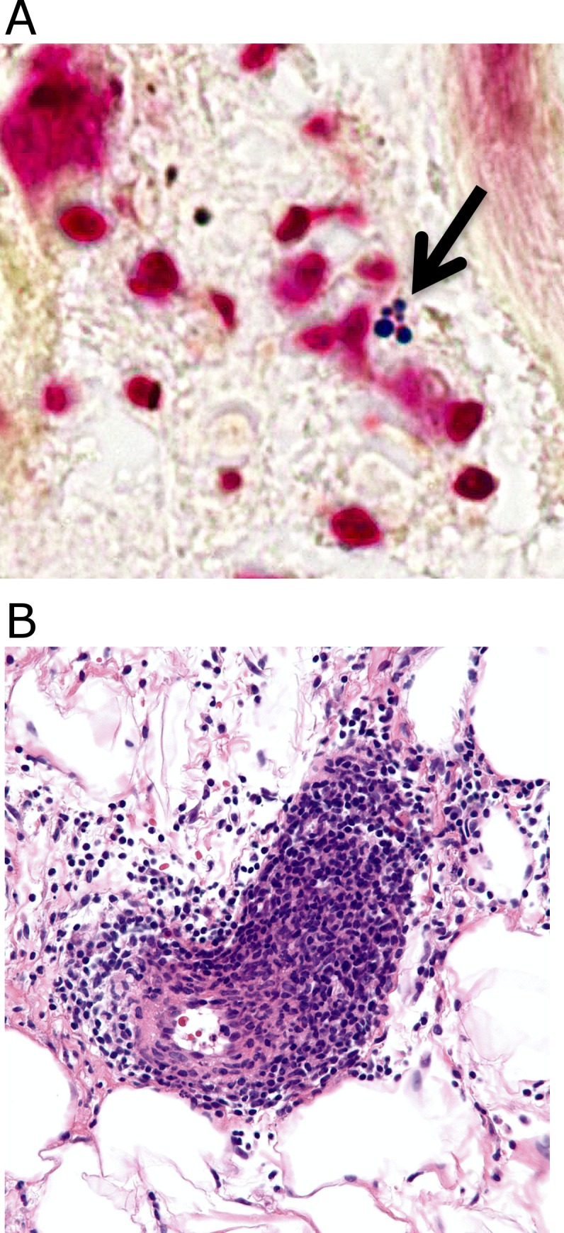
Fig 5. Photomicrographs from patients diagnosed clinically as having bacterial cellulitis.

Fig 5A: Culture-proven MRSA cellulitis with positive Gram stain. Note intracellular Gram-positive cocci in clusters. Fig 5B: Lymphocytic vasculitis misdiagnosed as cellulitis. Note abundant lymphocytes invading vessel wall.
